
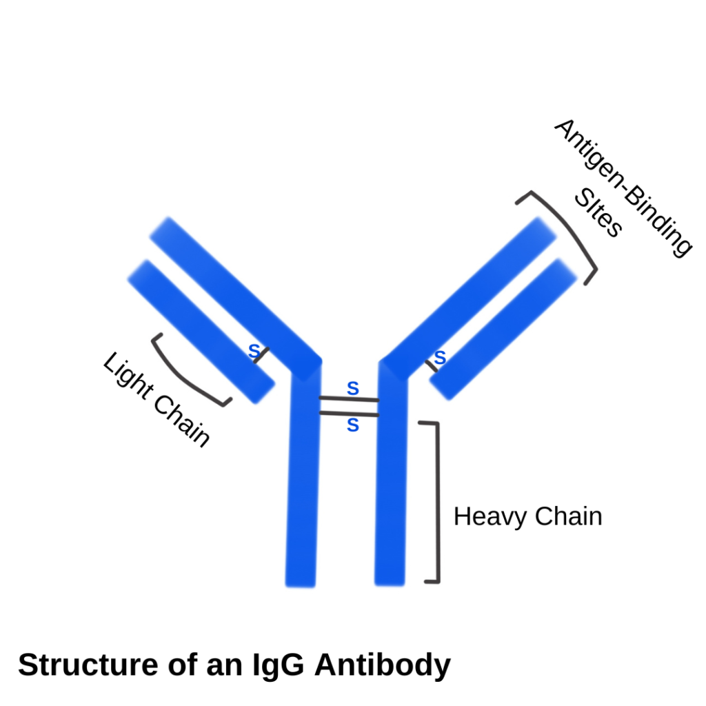
IgG antibodies are present in blood as tiny immunoglobulins under the radar. Due to their small size and less number of binding sites, they cannot cause visible agglutination such as IgM antibodies (present in ABO Blood Group System).
IgG antibodies are the main antibodies of Rh, Kell, Kidd, and Duffy blood group systems. The most common complication due to IgG antibodies is Rh antibody presence. Rh blood group system has five antibodies; D, E, C, e, c. Rh D antibody is of more clinical importance when it comes to blood transfusion reactions or in pregnancy situations. ABO blood group system mostly has IgM antibodies, which are easily visible in agglutination.
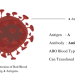
You may read the Simplifying ABO Blood Group System: Antigen, Antibody, and Transfusions to have a thorough understanding of blood groups; ABO and Rh antigens, antibodies.
Why Presence of IgG antibodies is Critical to Know?
The presence of IgG antibodies becomes critical to know when it comes to avoiding hemolytic transfusion reactions. The presence of IgG antibodies in the mother during pregnancy lethally causes hemolytic disease in the newborn (HDN). The issue with the identification of these antibodies is that they cannot cause visible agglutination as IgM does. They only bind to the antigens on red blood cells (sensitize them), but do not cause agglutination (visibly).
The most critical and common complication of Rh D antibody incompatibility is when an Rh-negative mother conceives a child who is Rh-positive. This condition gives rise to HDN, erythroblastosis fetalis, and hyperbilirubinemia in newborns.
Moreover, the presence of IgG antibodies also causes Autoimmune Hemolytic Anemia (AIHA). In AIHA, the body produces antibodies of IgG type against its antigens present in the body. In such conditions, it becomes significant to detect the presence of specific IgG antibodies in the blood plasma or on the surfaces of red blood cells. Further diagnostics would be set based on their presence and number in the body, as well as their type and class.
A Simple Test for The Identification of IgG Antibodies
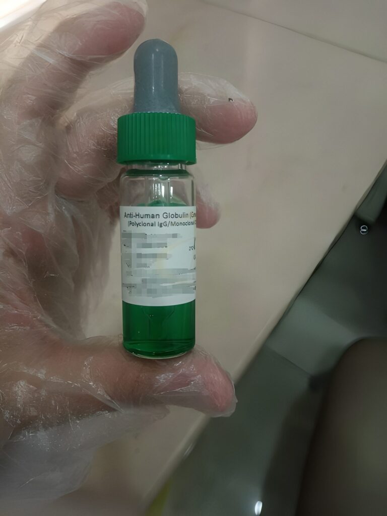
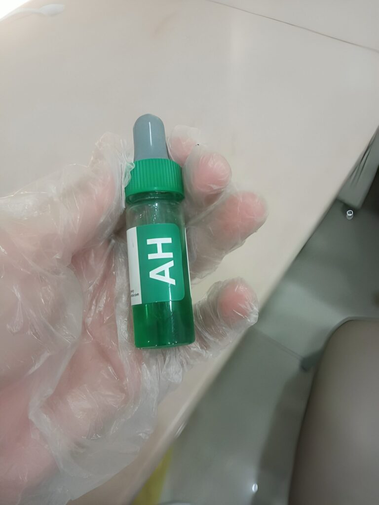
Coomb’s test is the most typical test for their identification. It is named after Dr. Robin Coombs, a British immunologist. He developed a method in the 1940s to detect the antibodies which are bind to the RBCs, and cannot produce visible agglutination. He developed a reagent to identify such (IgG) antibodies. This reagent is also called Coomb’s reagent, after him.
How Dr. Robin Coombs Developed the Coomb’s Reagent?
He found a way to obtain antibodies that work against the IgG antibodies in humans. He simply injected specific antibodies into the animals, most commonly rabbits and goats. The immune system of animals naturally produces antibodies against human antiglobulins (antibodies are also known as antiglobulins). After the natural time of the production of antibodies against human antibodies, he harvested and purified the blood plasma to obtain specific antibodies against those human antibodies that were injected in animals earlier. In this way, we got Anti-Human Globulin (AHG), another name for the Coomb’s reagent.
The development of Coomb’s reagent is a big milestone in controlling blood transfusion reactions, autoimmune hemolytic reactions, and maternal-fetal incompatibilities.
Dr. Coomb received the Lasker Award in 1972 for his contributions to medical science. However, he did not get a Nobel prize for his invention of Coomb’s reagent.
Types of Coomb’s Test for IgG Antibodies
There are two types of Coomb’s testing; Direct Antiglobulin Test (DAT) and Indirect Antiglobulin Test (IAT). There’s a striking difference between their sample collections and diagnosis.
We prefer DAT when we have to find the IgG antibodies present on the RBC surfaces. RBCs with IgG antibodies attached to them are known as sensitized cells. These are in vivo medium. IAT is done in vitro conditions. In this test, we detect antibodies that are present in the serum or plasma, not on the surface of RBCs. In IAT, we sensitize the IgG antibodies by incubating them with known red blood cells (more commonly O positive red blood cells) at 37 degrees Celsius with the LISS (Low Ionic Strength Solution) reagent. LISS minimizes the incubation period to 10 minutes. After incubation and centrifugation with the Coomb’s reagent, agglutination becomes visible.
Direct Antiglobulin Test (DAT)
While doing DAT, we take the samples of recipient red blood cells which have attached antibodies (in vivo sensitization). In DAT, one aim to detect antibodies attached with the RBCs. Following is the stepwise procedure for DAT testing;
- Take the blood sample of the recipient anti-coagulant EDTA tube. 3-4 ml blood is enough for DAT testing.
- Centrifuge the blood to separate blood cells and plasma for 10 mins at 3000-3500 rpm (centrifuge duration and speed depend upon the standard instructions of centrifuge machine).
- Take out the blood cells (sediment layer) in a separate tube.
- Wash the red blood cells three times with normal saline.
- After washing, make the 3% cell suspension with normal saline.
- Add one drop of 3% cell suspension of normal saline in a tube for Coomb’s testing.
- Add two drops of Coomb’s Reagent in the above tube (to which we have added one drop of 3% red cell suspension).
- Mix the ingredients and centrifuge the sample for 15 seconds and 3500 rpm.
- Check for agglutination.
What does the Coomb’s Reagent Do?
Coomb’s reagent makes the agglutination visible because of antibody against antibody reaction. Coomb’s reagent binds to the IgG antibodies present on the surface of RBCs in DAT. It holds the chains of IgG antibodies together, causing their clumping that makes agglutination visible. Agglutination confirms the presence of IgG antibodies present in vivo.
Indirect Antiglobulin Test (IAT)
IAT has an application to detect antibodies freely present in plasma or serum. After centrifugation, the whole blood, red blood cells, and plasma become separate in the form of layers. We do IAT on the plasma or the upper layer after centrifugation. Following is the step-by-step procedure of IAT;
- Take two drops of the recipient’s serum in a test tube (after centrifugation).
- Add one drop of 3% O-positive red blood cell suspension to this tube.
Note: We use O-positive red blood red blood cells in the Indirect Antiglobulin Test (IAT) to let the antibodies in the recipient’s serum (if any) attach to the surface of O-positive red blood cells. Furthermore, the specificity of using O-positive red blood cells for IAT is due to the absence of A and B antigens on the red blood cells of this blood type. O-positive red blood cells only have Rh D antigens. Rh blood system has IgG antibodies for which we are doing Coomb’s testing. In a nutshell, using O-positive red blood cells in this test would simplify things. If agglutination occurs, it would directly indicate the presence of IgG antibodies (antibodies against Rh D antigens or other minor blood group systems). These antibodies are known as alloantibodies.
Alloantibodies are formed in the body against non-self red blood cell antigens. They form after a blood transfusion, or pregnancy, etc. The presence of alloantibodies in the recipient serum is a potential threat for post-transfusion reaction as they tend to react with donor red blood cell antigens.
- Incubate the above-prepared tube for 10 mins at 37 degrees Celsius with LISS reagent.
- After incubation, wash the cells 3 times with saline.
- After decanting the supernatant by 3rd saline washing, add two drops of Coomb’s reagent to it.
- Centrifuge the remaining sample for 15 mins at 3500 rpm.
- Check for visible agglutination.